Mitral Stenosis: A Crucial Concept for Every Medical Student
Navigating the complexities of cardiology can feel overwhelming, but some conditions demand your focused attention. Mitral stenosis is one such topic that every medical student needs to have firmly in their grasp. Understanding its pathophysiology, causes, symptoms, and management is fundamental for your academic success and future clinical practice. This article breaks down mitral stenosis, offering a clear and concise overview to aid your studies. Remember, active recall is key to mastering medical concepts, and you can transform your own notes into powerful study tools with Quizflex AI.
The Core of the Issue: What is Mitral Stenosis?
At its heart, mitral stenosis is a condition characterized by the narrowing of the mitral valve. This valve, situated between your left atrium and left ventricle, plays a critical role in ensuring unidirectional blood flow. When it's stenotic, it means the valve leaflets have become thickened and stiff, preventing them from opening fully.
During diastole, the period when the heart relaxes and fills with blood, the mitral valve should open wide to allow oxygenated blood from the left atrium to flow into the left ventricle. In mitral stenosis, this opening is restricted. This restriction leads to:
- Reduced forward blood flow: Less blood can enter the left ventricle, impacting the amount of blood pumped to the rest of the body.
- Pressure gradient: A difference in pressure develops between the left atrium and the left ventricle because blood struggles to pass through the narrowed valve. This elevated pressure in the left atrium can have significant downstream effects.
Unpacking the Causes: Where Does Mitral Stenosis Originate?
While various factors can contribute to mitral stenosis, three primary causes stand out and are essential for you to remember:
-
Rheumatic Fever: This is the most common cause of mitral stenosis globally, particularly prevalent in developing regions. Rheumatic fever is an inflammatory disease that can occur after an untreated streptococcal infection (like strep throat). The inflammation can damage heart valves, including the mitral valve, leading to scarring and thickening over time. This chronic damage is why understanding the sequelae of common infections is so vital in medicine.
-
Age-Related Calcification: In developed countries, as populations age, calcification of the mitral valve becomes a more frequent contributor to stenosis. Similar to how arteries can harden with age, the mitral valve can also accumulate calcium deposits. These deposits can stiffen the valve leaflets, impeding their proper function. This highlights the importance of considering age as a factor in cardiovascular disease presentation.
-
Congenital Defects: Though less common, mitral stenosis can be present from birth. These are congenital heart defects where the mitral valve may not have formed correctly, leading to narrowing from infancy. Recognizing congenital conditions is crucial for pediatric cardiology and for understanding the full spectrum of heart disease.
Recognizing the Signs: Key Symptoms of Mitral Stenosis
The symptoms of mitral stenosis often develop gradually and can be insidious. They are a direct consequence of the impaired blood flow and the increased pressure within the left atrium. For learners, recognizing the hallmark symptoms is paramount for diagnosis:
-
Dyspnea (Shortness of Breath): This is often the hallmark symptom of mitral stenosis. Initially, you might notice it primarily during exertion (exertional dyspnea). As the condition progresses, shortness of breath can occur even at rest. This occurs because the elevated pressure in the left atrium backs up into the pulmonary veins, causing fluid to accumulate in the lungs (pulmonary congestion).
-
Fatigue: Reduced cardiac output means less oxygenated blood is delivered to the body's tissues, leading to generalized fatigue and a feeling of being easily tired.
-
Cough: A persistent cough, especially one that may be worse when lying down, can be a sign of pulmonary congestion. In some cases, it might even produce frothy sputum, indicating significant fluid buildup in the lungs.
-
Chest Pain: While not as common as dyspnea, some individuals with mitral stenosis may experience chest pain. The exact mechanism can vary but may be related to increased workload on the heart or pulmonary hypertension.
-
Hemoptysis (Coughing Blood): This is a less common but clinically significant symptom. It occurs when the high pressure in the pulmonary veins causes small blood vessels in the lungs to rupture. It's a red flag that indicates severe mitral stenosis and significant pulmonary congestion.
Navigating Treatment: Tiers of Intervention
The management of mitral stenosis depends on the severity of the condition, the patient's symptoms, and their overall health. Treatment strategies are typically tiered, starting with less invasive options and progressing to more definitive interventions if necessary:
-
Medications:
- Diuretics: These medications are used to help manage fluid overload and reduce congestion in the lungs, thereby alleviating shortness of breath.
- Beta-blockers: If the patient develops atrial fibrillation (a common complication, discussed later), beta-blockers are often prescribed to help control the heart rate.
- Anticoagulants: For patients with atrial fibrillation, anticoagulants (blood thinners) are crucial to prevent the formation of blood clots in the enlarged left atrium. These clots can then travel to the brain, causing a stroke.
-
Percutaneous Balloon Mitral Valvotomy (PBMV): This is a minimally invasive procedure often considered a first-line treatment, especially in younger patients who have pliable valve leaflets. A catheter is inserted into a vein, guided to the heart, and a balloon is inflated within the narrowed mitral valve to widen it.
-
Valve Repair or Replacement: For patients with severe, symptomatic mitral stenosis, or when PBMV is not a suitable option, surgical intervention may be necessary. This can involve repairing the damaged valve or, in more severe cases, replacing it with a mechanical or biological prosthetic valve.
Clinical Pearls for Deeper Understanding
To truly master mitral stenosis, internalize these critical clinical pearls:
-
Auscultation Findings: When listening to the heart with a stethoscope, pay close attention to the characteristic sounds. In mitral stenosis, you'll often hear an opening snap followed by a low-pitched diastolic rumble best heard at the apex of the heart. These sounds are direct evidence of the stenotic valve's movement during the filling phase of the cardiac cycle.
-
Association with Atrial Fibrillation: Mitral stenosis significantly increases the risk of developing atrial fibrillation (AFib). The enlarged left atrium, a consequence of the pressure buildup, can become electrically unstable, leading to irregular and rapid heartbeats.
-
Thromboembolism Risk: The enlargement of the left atrium due to mitral stenosis creates a stagnant area where blood can pool and clot. This makes thromboembolism (the formation and travel of blood clots) a serious concern. This is precisely why anticoagulation is so vital in patients with AFib secondary to mitral stenosis.
-
Progression to Pulmonary Hypertension and Right Heart Failure: Chronic obstruction of blood flow from the left atrium to the left ventricle can lead to a backup of pressure into the pulmonary circulation. This can cause pulmonary hypertension (high blood pressure in the arteries of the lungs). Over time, the right side of the heart has to work harder to pump blood against this increased resistance, potentially leading to right heart failure.
Visualizing the Concept
To solidify your understanding, consider this visual aid:
Why This Matters for Your Learning Journey
Short, impactful visuals like the one above are excellent for getting a quick overview of a topic. They serve as a fantastic starting point before you dive deep into your textbooks or lecture notes. However, to truly make this knowledge stick, you need to engage in active recall. This means actively testing yourself on the material.
Transform Your Notes into Powerful Quizzes
Are you studying cardiology, pathology, or preparing for exams like USMLE Step 1 or Step 2? The most effective way to learn is by turning your own study materials into interactive quizzes. Quizflex AI is designed specifically for medical students. Simply upload your notes, PDFs, or even lecture transcripts, and our AI will generate a variety of question formats, including multiple-choice, fill-in-the-blank, and short-answer questions. You can even get AI-explained answers to deepen your comprehension.
Stop passively rereading and start actively testing yourself. Make your study sessions more efficient and effective with Quizflex AI.
Related: Blog home · Pricing · AI quiz generator
Mitral stenosis — every med student needs to memorize this. 🫀
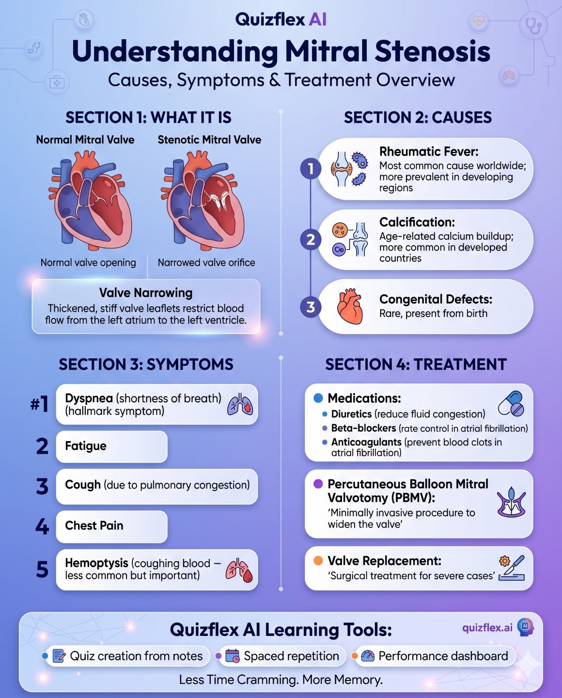
Mitral stenosis is the narrowing of the mitral valve, restricting blood flow from the left atrium to the left ventricle. Here's the complete clinical picture:
🫁 WHAT IT IS:
Thickened, stiff valve leaflets that don't open fully — restricting forward blood flow during diastole and creating a pressure gradient.
⚡ TOP 3 CAUSES:
1. Rheumatic fever — most common cause worldwide, especially in developing regions
2. Age-related calcification — common in older adults in developed countries
3. Congenital defects — rare, present from birth
🚨 KEY SYMPTOMS:
1. Dyspnea — exertional shortness of breath (the hallmark)
2. Fatigue
3. Cough — due to pulmonary congestion
4. Chest pain
5. Hemoptysis — coughing blood, less common but clinically important in severe cases
🩺 TREATMENT TIERS:
- Medications:
— Diuretics (reduce fluid congestion)
— Beta-blockers (rate control in atrial fibrillation)
— Anticoagulants (prevent blood clots if AFib present)
- PBMV (Percutaneous Balloon Mitral Valvotomy) — minimally invasive, often first-line in younger patients
- Valve repair or replacement — surgical option for severe symptomatic cases
🔑 Clinical pearls:
✅ Listen for the OPENING SNAP and low-pitched diastolic rumble at the apex
✅ Often associated with atrial fibrillation
✅ Left atrial enlargement increases risk of thromboembolism — hence anticoagulation
✅ Severe cases can lead to pulmonary hypertension and right heart failure
📚 Studying cardiology, pathology, or USMLE Step 1/2? Drop your notes into Quizflex AI for instant flashcards, MCQs, and AI-explained answers — built for med students.
🔗 quizflex.ai
Save this for your cardiology block 📌
#MedSchool #Cardiology #USMLE #Step1 #Step2 #MedStudent #Nursing #PreMed #StudyGram #StudentLife #ExamPrep #QuizflexAI #MitralStenosis #HeartConditions #MedicalEducation #InternalMedicine #StudyTips #MedTwitter
Topics covered
- Quizflex
- Infographic
- Study tips
- Medschool
- Cardiology
- Usmle
- Step1
- Step2
- Medstudent
- Nursing
Frequently asked questions
What is Mitral stenosis - every med student needs to memorize this. 🫀 Mitral stenosis is the narrowing of t?
How do I study Mitral stenosis - every med student needs to memorize this. 🫀 Mitral stenosis is the narrowing of t effectively?
What is the best AI quiz tool for Mitral stenosis - every med student needs to memorize this. 🫀 Mitral stenosis is the narrowing of t?
How can I practice Mitral stenosis - every med student needs to memorize this. 🫀 Mitral stenosis is the narrowing of t with QuizFlex AI?
Is QuizFlex AI free for students and teachers?
Stay in the Loop
Get weekly tips on AI-powered learning, quiz strategies, and product updates. No spam, unsubscribe anytime.
POV: it's 11pm, exam's tomorrow, and you've got 80 pages of notes you haven't touched 💀 stop. breat
Studying for 5 hours straight doesn't make you a better student. ⏱️ It usually means you'll forget m
Related Articles
Continue learning with these related posts
Right now, 1 billion germs are trying to infect you. 🦠 And right now, your body is winning. Every.
Right now, 1 billion germs are trying to infect you. 🦠 And right now, your body is winning. Every. Single. Day. Here's how your immune system actually fig…
Anxiety shrinks WORKING memory. Breathe first - then ONE question. - Small steps lower the activa
Anxiety shrinks WORKING memory. Breathe first - then ONE question. - Small steps lower the activation barrier How Quizflex AI helps you practice and remember.
Every human embryo starts out exactly the same.
Every human embryo starts out exactly the same. - concepts, study ideas, and practicing with Quizflex AI.